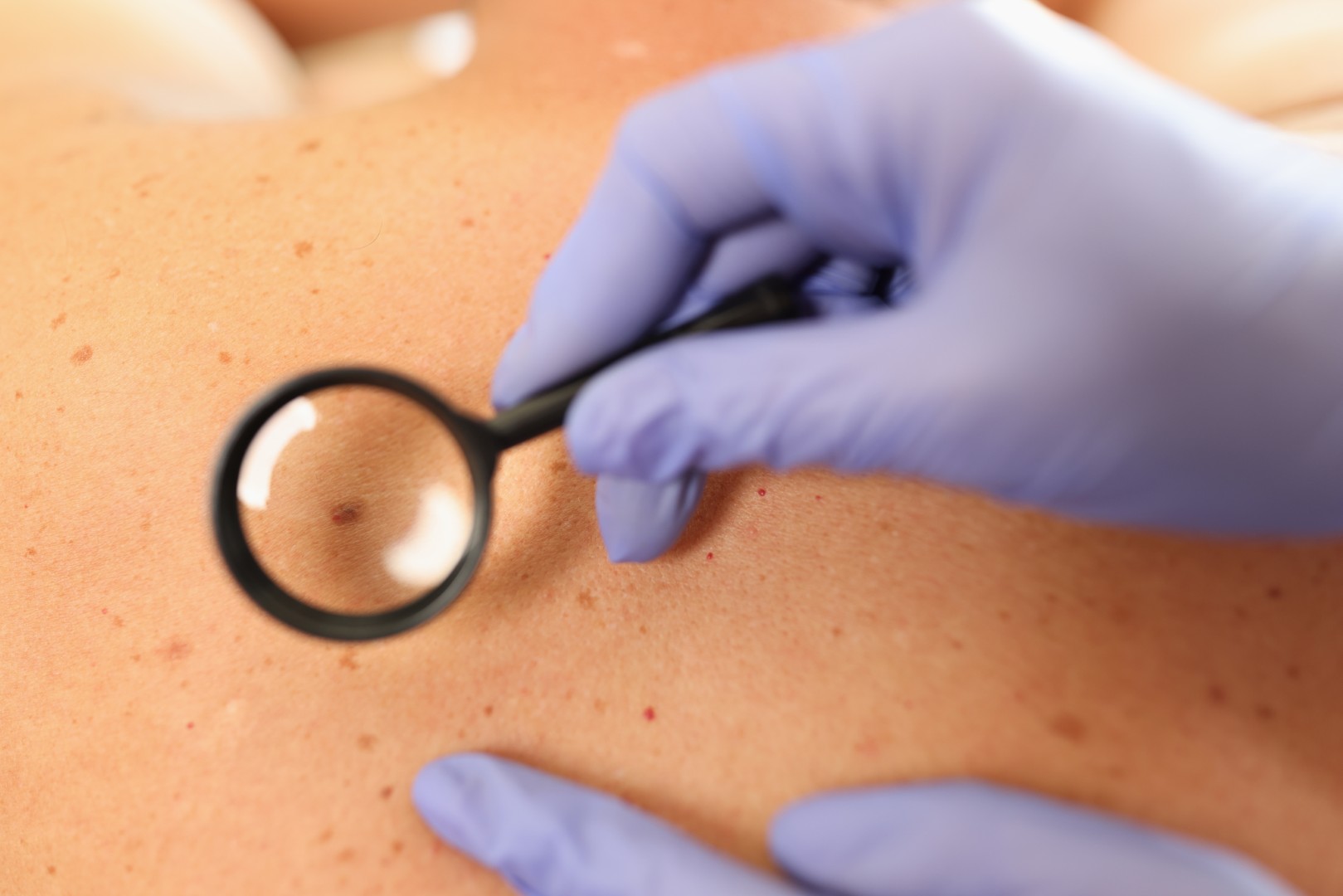


The use of dermoscopy and digital nevus mapping significantly contributes to the early diagnosis of melanoma and helps reduce unnecessary surgical removal of benign skin lesions, provided that the procedure is performed by a well-trained and experienced dermatologist skilled in this technique.
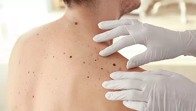
Digital mole mapping is the most reliable method for early diagnosis of skin cancer. A special camera records the moles (olives) scattered throughout the body and classifies them as suspicious, benign or malignant. The images are stored and used in the re-examination of the moles, allowing us to monitor the evolution of each mole with an accurate comparison of the previous with the recent image. This is a painless examination, without radiation and suitable for all ages.
Digital mole mapping is necessary for people with:
- multiple-dysplastic nevi,
- personal or family history of melanoma,
- weakened immune system,
- history of sunburn.
Common Questions
I have about 50 olives on my body. Do I have to map them all? If not, who are the candidates?
Of course, we do not “map” all of our olive trees. However, our dermatologist must examine them all dermatoscopically. The question of which ones we will analyze and record digitally will be answered by our doctor. Dermoscopic and clinical criteria are not always those seemingly apparent to the naked eye.
So from what age can someone take this test?
From birth, if necessary. It is a completely safe test. In children, it is used for detailed monitoring of congenital moles.
Does mapping only help us diagnose melanoma or other forms of skin cancer?
In addition to melanoma, there are other forms of skin cancer, which are more common and not as life-threatening as melanoma. Very often, during the examination—especially in middle-aged and older individuals—other forms of skin cancer can be detected and diagnosed, such as basal cell carcinoma or squamous cell carcinoma.